Back Of Neck Anatomy / : Watch cervical muscle anatomy animation. In addition, in this region we also find the major cranial and spinal nerves that connect the central nervous system to the organs, skin, and muscles of the head and neck. Neck muscles can be strained from poor posture — whether it's leaning over your computer or hunching over your workbench. It consists of seven vertebrae. An area called the occiput. In this video, i walk you through a basic approach to drawing the neck and upper back muscles.
The crossword solver found 20 answers to the back of the neck crossword clue. This is a more stylized study and not meant to be entirely cor. Remember, while c1 and c2 allow tremendous ranges of neck movement, they are supporting your head too. Anatomy of the back 12 photos of the anatomy of the back anatomy lungs back view, anatomy of a red back spider, anatomy of the back and kidneys, anatomy of the back neck muscles, anatomy of the human body back muscles, human anatomy, anatomy lungs back view, anatomy of a red back spider, anatomy of … Contains glands ( thyroid , parathyroid, and thymus ), the larynx , pharynx and trachea.
Muscles of the Neck • Bodybuilding Wizard from bodybuilding-wizard.com The majority of these nerves control the functions of the upper extremities and allow you to feel your arms, shoulder, and back of your head. The four parathyroid glands are situated upon the back surface of the thyroid gland. Rarely, neck pain can be a symptom of a more serious problem. Causes of neck pain and how to manage the pain in basic terms, the neck (cervical spine) joins the shoulders and chest to the head. Jugularis posterior) begins in the occipital region and returns the blood from the skin and superficial muscles in the upper and back part of the neck, lying between the splenius and trapezius. The structures of the human neck are anatomically grouped into four compartments; The back anatomy includes the latissimus dorsi, trapezius, erector spinae, rhomboid, and the teres major. Within these compartments, the neck houses the cervical vertebrae and cervical part of the spinal cord, upper parts of.
The structures of the human neck are anatomically grouped into four compartments;
The back of the neck is mostly comprised of muscles, as well as the spine. Causes of neck pain and how to manage the pain in basic terms, the neck (cervical spine) joins the shoulders and chest to the head. This is a more stylized study and not meant to be entirely cor. After arising from the common carotid artery, it travels up the neck, passing posteriorly to the mandibular neck and anteriorly to the lobule of the ear. It consists of seven vertebrae. Click the answer to find similar crossword clues. Muscle head anatomy vocal organ diagram female neck anatomy neck wireframe head neck human anatomy head artery anatomy face pharynx vector neck degree head anatomy 3d. The external carotid artery supplies the areas of the head and neck external to the cranium. Neck anatomy explained the neck begins at the base of the skull and connects to the thoracic spine (the upper back). These muscles give the sides of the neck their. Anatomy of the back 12 photos of the anatomy of the back anatomy lungs back view, anatomy of a red back spider, anatomy of the back and kidneys, anatomy of the back neck muscles, anatomy of the human body back muscles, human anatomy, anatomy lungs back view, anatomy of a red back spider, anatomy of … The top of the cervical spine connects to the skull, and the bottom connects to the upper back at about shoulder level. Within these compartments, the neck houses the cervical vertebrae and cervical part of the spinal cord, upper parts of.
Remember, while c1 and c2 allow tremendous ranges of neck movement, they are supporting your head too. After arising from the common carotid artery, it travels up the neck, passing posteriorly to the mandibular neck and anteriorly to the lobule of the ear. The neurocranium (cranial vault) and the viscerocranium (facial skeleton). Anatomy of the back 12 photos of the anatomy of the back anatomy lungs back view, anatomy of a red back spider, anatomy of the back and kidneys, anatomy of the back neck muscles, anatomy of the human body back muscles, human anatomy, anatomy lungs back view, anatomy of a red back spider, anatomy of … The structures of the human neck are anatomically grouped into four compartments;
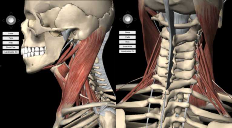
Neck Muscles Anatomy - Posterior Triangle, Prevertebral ... from i.ytimg.com Neck pain is a common complaint. Within these compartments, the neck houses the cervical vertebrae and cervical part of the spinal cord, upper parts of. On this page, you'll learn about each of these muscles, their locations and functional anatomy. The external carotid artery supplies the areas of the head and neck external to the cranium. The larynx is located where the pharynx, the back of the mouth and nasal cavity, divides into the trachea (the tube that carries air to the lungs) and the esophagus (the tube that carries food to. The top of the cervical spine connects to the skull, and the bottom connects to the upper back at about shoulder level. The crossword solver found 20 answers to the back of the neck crossword clue. The posterior triangle of the neck is an anatomical area located in the lateral aspect of the neck.
The neck is the part of the body on many vertebrates that connects the head with the torso and provides the mobility and movements of the head.
Contains glands ( thyroid , parathyroid, and thymus ), the larynx , pharynx and trachea. The occipital bone surrounds a large opening known as the foramen magnum. Function of the back muscles there are several individual muscles within the back anatomy, and it's important to take a quick look at all of Contains cervical vertebrae and postural muscles. The neurocranium (cranial vault) and the viscerocranium (facial skeleton). Jugularis posterior) begins in the occipital region and returns the blood from the skin and superficial muscles in the upper and back part of the neck, lying between the splenius and trapezius. The majority of these nerves control the functions of the upper extremities and allow you to feel your arms, shoulder, and back of your head. It is made up of bones, discs, muscles, ligaments, nerves and tendons. The posterior triangle of the neck is an anatomical area located in the lateral aspect of the neck. Rarely, neck pain can be a symptom of a more serious problem. Causes of neck pain and how to manage the pain in basic terms, the neck (cervical spine) joins the shoulders and chest to the head. The skull is a strong, bony capsule that rests on the neck and encloses the brain. Watch cervical muscle anatomy animation
See more ideas about back pain, spine health, spine problems. Neck pain is a common complaint. Neck anatomy explained the neck begins at the base of the skull and connects to the thoracic spine (the upper back). Cervical spine anatomy video the cervical spine has 7 stacked bones called vertebrae, labeled c1 through c7. Think of it like a jigsaw puzzle, all the pieces fit in together and are required to get the full picture as to how it works.
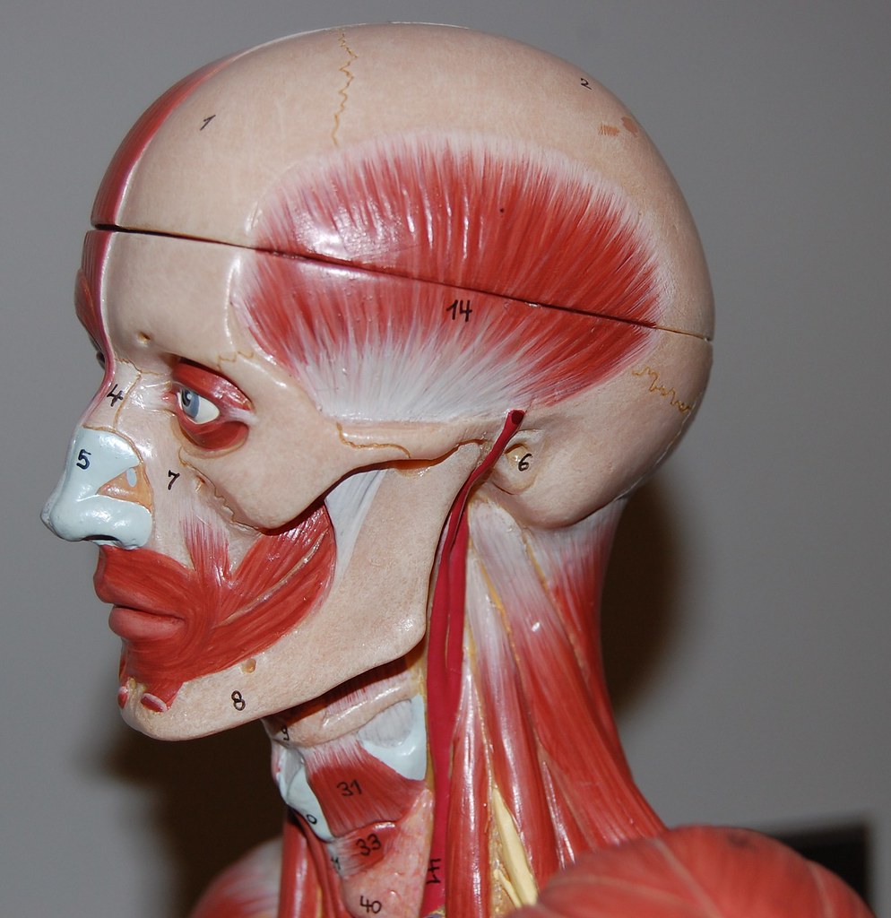
Muscles of the head and neck, left lateral view | Rob ... from c1.staticflickr.com This is a more stylized study and not meant to be entirely cor. Enter the answer length or the answer pattern to get better results. The majority of these nerves control the functions of the upper extremities and allow you to feel your arms, shoulder, and back of your head. These muscles give the sides of the neck their. This muscle is controlled by the third and fourth cervical nerves (c3, c4). It consists of seven vertebrae. Think of it like a jigsaw puzzle, all the pieces fit in together and are required to get the full picture as to how it works. It consists of two major parts:
Contains glands ( thyroid , parathyroid, and thymus ), the larynx , pharynx and trachea.
The occipital bone surrounds a large opening known as the foramen magnum. On this page, you'll learn about each of these muscles, their locations and functional anatomy. They move the head in every direction, pulling the skull and jaw towards the shoulders, spine, and scapula. Within these compartments, the neck houses the cervical vertebrae and cervical part of the spinal cord, upper parts of. The posterior external jugular vein (v. The vertebral bodies are round shapes. Enter the answer length or the answer pattern to get better results. The crossword solver found 20 answers to the back of the neck crossword clue. The neck is essentially a passageway for air, food, liquids, blood, and more to travel between the head and the rest of the body, through structures such as blood vessels, nerves, and lymph nodes, as well as the larynx, trachea, and esophagus. The cervical spine protects the nerves connecting to the brain, allowing the head to move freely while supporting its weight. Osteoarthritis also is a common cause of neck pain. Muscle head anatomy vocal organ diagram female neck anatomy neck wireframe head neck human anatomy head artery anatomy face pharynx vector neck degree head anatomy 3d. After arising from the common carotid artery, it travels up the neck, passing posteriorly to the mandibular neck and anteriorly to the lobule of the ear.


0 Comments